Brain-eating amoeba is as deadly as the name sounds. It is also known as Naegleria fowleri. It is a single-celled organism that was first discovered in 1965 and is usually found in warm freshwater bodies or untreated and contaminated waters.
It causes a rare disease that destroys the human brain by feeding on it. The deadly micro-organism can even lead to death. Know all about this amoeba in detail and how to treat the disease caused by it.
What Is Brain-Eating Amoeba And Where Does It Live?
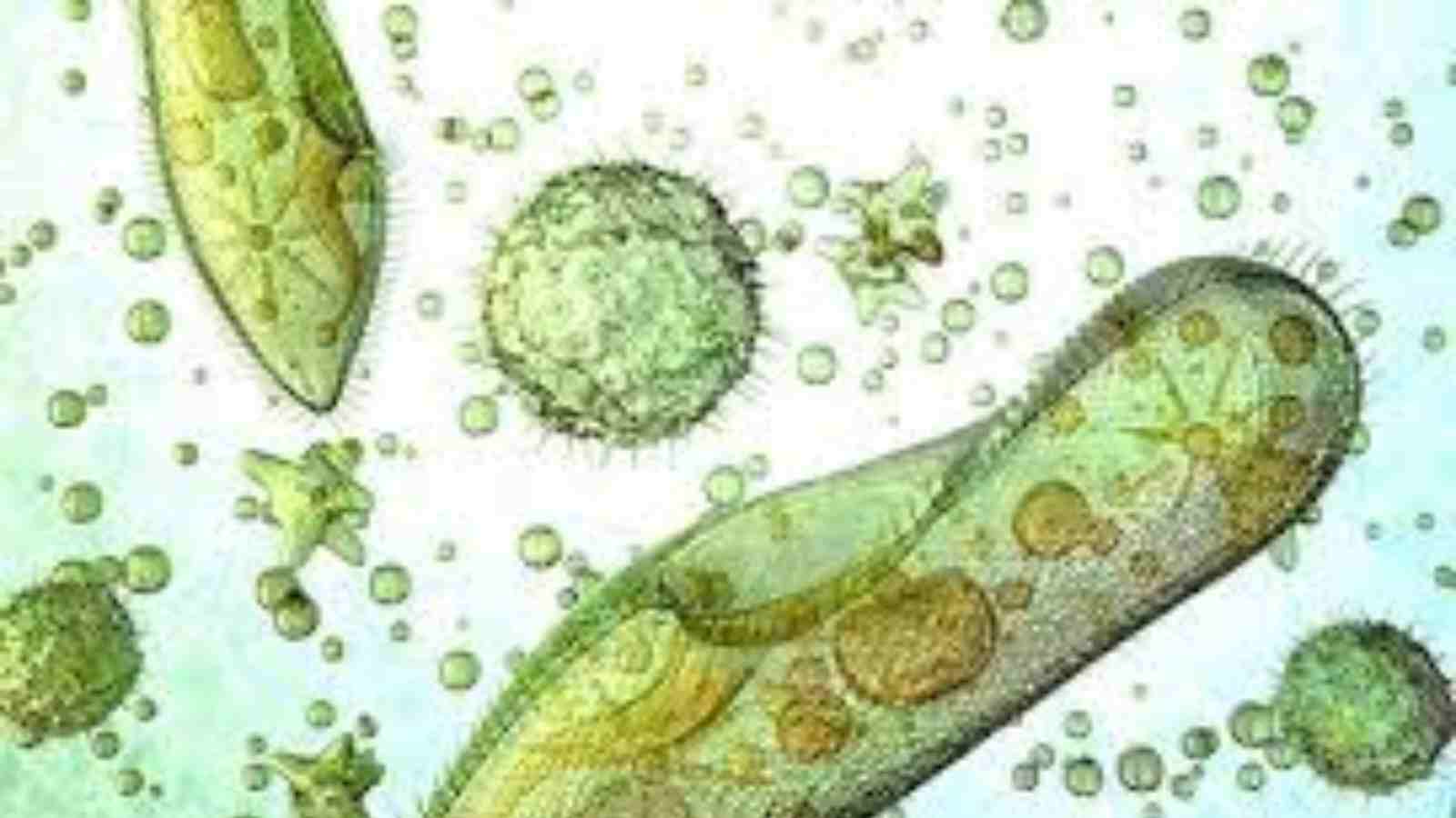
Naegleria fowleri, also known as brain-eating amoeba, belongs to the phylum Percolozoa, which is not a true amoeba. It is named after an Australian pathologist, Malcolm Fowler. It is a single-celled, heat-loving, free-living, bacteria-eating, and thermophilic microorganism. These are excavated and live in soil and water. The amoeba is sensitive to acid, and drying and cannot live in seawater. It grows best at temperatures up to 46 °C. It lives in warm lakes, ponds, mud puddles, and rock pits. They are even found in warm, slow-flowing rivers, especially those with low water levels. Untreated swimming pools, spas, well water, and municipal water are also suitable for them. It cannot survive in water that undergoes proper treatment.
Other places where Naegleria fowleri lives are hot springs, and thermally polluted water which include water from power plants and aquariums. Soil including indoor dust is also very suitable for them. Many cases of it have been reported from Southern and South Western states of the U.S. Especially, many cases came from Florida and Texas. Outside the States, cases were reported from Pakistan as well.
Life Cycle
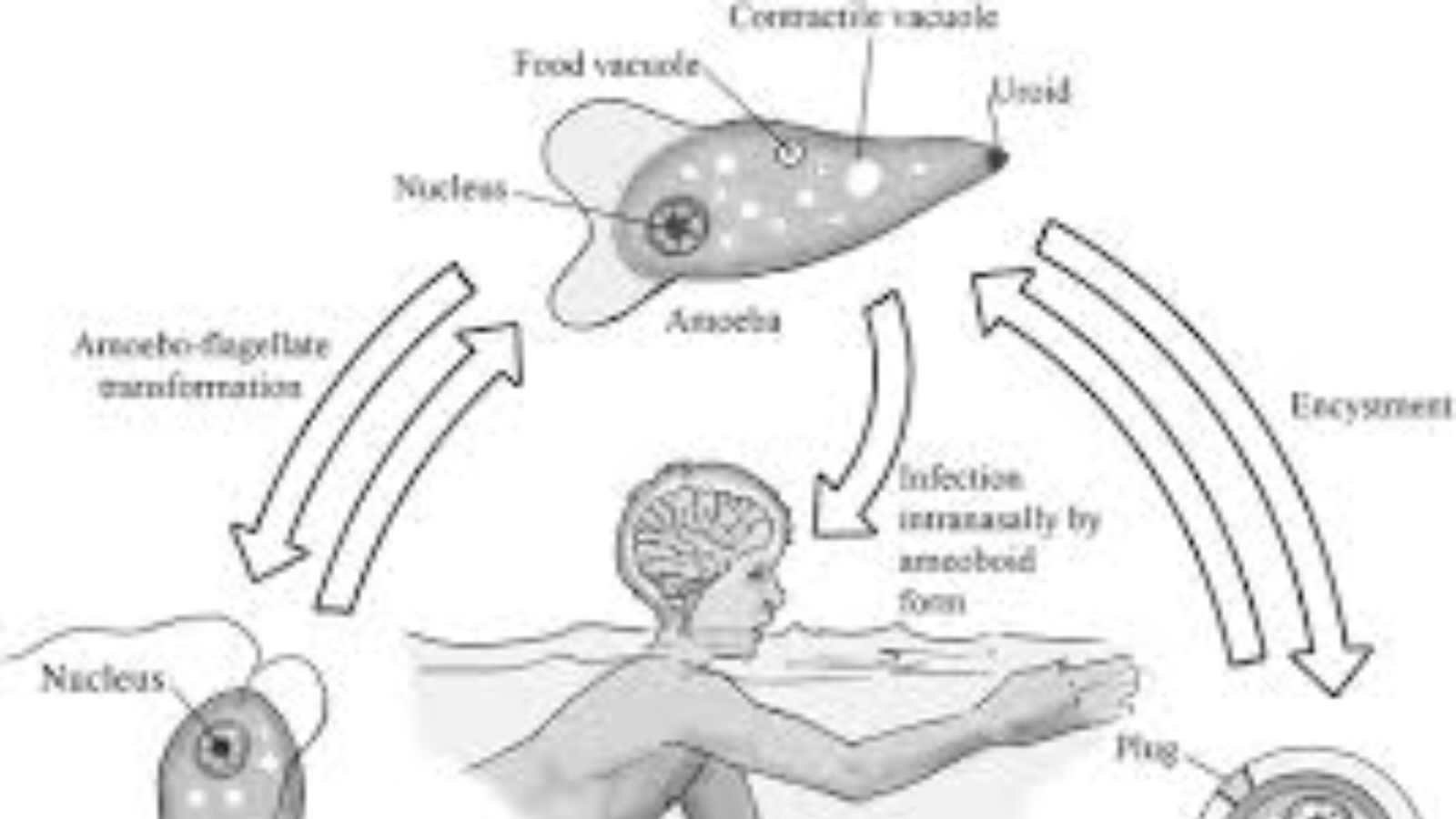
It goes through three stages in its life cycle. They are namely – cyst, flagellate, and trophozoite. The trophozoite change to form cysts which can survive even in harsh environmental conditions. It is a spherical, single-layered, smooth wall which encloses the single nucleus. When there is a suitable condition the amoeba escapes through a pore present in the middle. Encystation occurs below 10 °C.
The flagellate stage is pear-shaped and has two flagellas. It is not the infective form of the organism, it just roams around. However, they may go into the nasal cavity during swimming or diving by inhalation. It then transforms into trophozoite and causes infections.
The trophozoite stage is the stage that is infective to man. It can actively eat and reproduce by binary fission. They attach to the olfactory epithelium, where it follows the olfactory cell axon through the cribriform plate to reach the brain. They travel using pseudopodia. In tissues, the trophozoites phagocytize red blood cells and destroy tissue by releasing cytolytic substances. Animals and humans act as accidental hosts for the trophozoites.
Pathogenicity
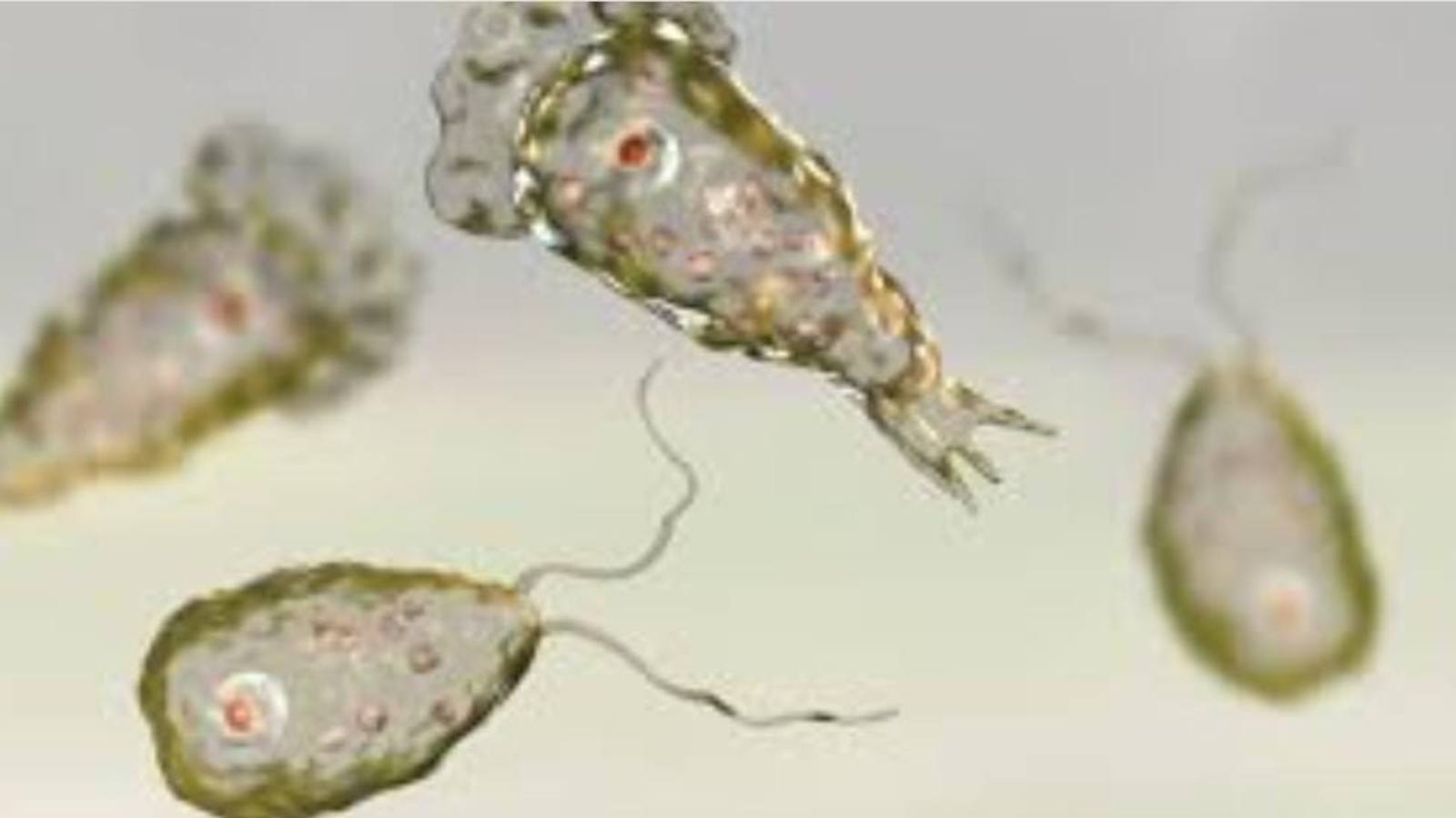
N. fowleri causes a lethal infection to the brain known as naegleriasis. It is also known as primary amoebic meningoencephalitis (PAM). It enters by inhalation through the nose and travels by nasal and olfactory nerve to the brain. Inhalation is the only means of its entry, it cannot enter by swallowing of the contaminated water. The amoeba normally eats bacteria but in humans, it consumes astrocytes and neurons present.
Symptoms tend to appear one to nine days after the entry of flagellates. Early symptoms are headache, fever, nausea, vomiting, loss of appetite, altered mental state, coma, drooping eyelid, blurred vision, and loss of the sense of taste. Late symptoms include stiff neck, confusion, lack of attention, loss of balance, seizures, and hallucinations. Once the symptoms start to occur, death usually follows within two weeks.
Diagnosis And Treatment
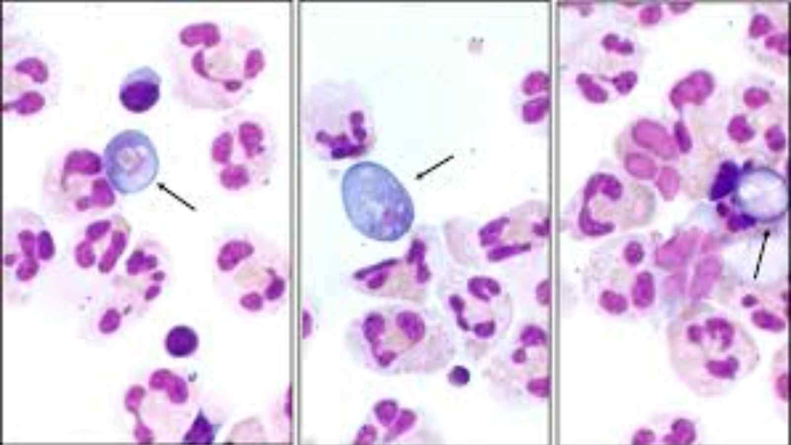
Samples collected to diagnose this amoeba are cerebrospinal fluid, biopsy, or tissue specimens. As this infection is very rare there are not many rapid tests available to detect it. Amoeba cultures and real-time PCR studies are some diagnostic tools that are available in very few laboratories. The treatment includes the antifungal drug amphotericin B, which inhibits it by binding to the cell membrane leading to cell membrane disruption and finally death.
However, even with this, the fatality rate remains above 95%. Miltefosine – an antiparasitic drug can also be considered as a treatment measure. It inhibits the PI3K/Akt/mTOR pathway which is responsible for cell survival.
Read More: Hailey Bieber Shares Husband Justin Bieber’s Health Update Post Ramsay Hunt Syndrome Diagnosis

